Routine Eye Examinations
There are number of tests that your doctor may perform during your normal routine eye exam to ensure that your vision is at its best. Some of these tests include:

Visual Field Screening
A Visual Field Test will allow your doctor to see the full horizontal and vertical range of what are are able to see peripherally. This enables the doctor to determine the potential presences of blind spots (scotomas), which could indicate eye diseases.
Visual Field Screenings are performed routinely at your exam with an automated computer screening instrument. If an eye disease is suspected, further comprehensive, more formal tests may be required.
This is the "Clicker Test".

Autorefractor Testing
An autorefractor is sometimes used by doctors to determine a patient's prescription. A chin and forehead rest will help stabilize your head while you look at a pinpoint of light—the hot air balloon. It is used to evaluate the way your retina focuses an image.
The Visual Field Screening and Autorefractor Test is usually performed pre-exam by one of our trained Opticians.

Retinoscopy
A retinoscopy test will occasionally be peformed early in the eye exam so that the doctor can determine an approximate prescription. With the room's lights dim, you will be asked to look through a machine and focus on a large target (usually the big "E" on the eye chart). The doctor will shine a light in your eye, while selecting different lenses. Based on how the light reflects on the retina, your refractive error will be calculated.
Autorefractive technology has allowed this reading to be obtained now by a computerized scan.

Refraction Assessment
The Refraction Assessment determines the eyeglass prescription. It helps the doctor determine the most accurate corrective lenses that will give you the best possible vision. You will be asked to look through a Phoroptor—a mask-like device that contains different lenses—which will help determine the best combination to give you the sharpest vision.
Which looks better...one...or two?

Visual Acuity Test
A Visual Acuity Test is a part of an eye exam performed to determine the smallest letters a patient can read on a standardized chart that is 20 feet away. It is done without the aid of contacts or glasses, and only one eye is tested at a time. With one eye covered, you will be asked to read the smalled line of letters that you can read on that chart.
Visual Acuity is expressed as a fraction, with the top number representing the distance one is standing from the chart—20 feet—and the bottom number representing the distance at which a person with normal eyesight could read the same line that was read correctly.
Normal vision is, thus, 20/20. If your acuity is 20/40, this indicates that what you can correctly read at 20 feet can be read by a person with normal vision from 40 feet away.
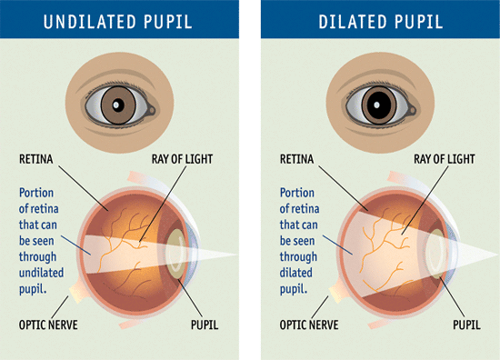
Dilation (A.K.A. The Drops)
Dilating eye drops are routinely used on all patients. They are used to enlarge the pupil and allow the doctor to see more of the Retina, including the periphery, when evaluating patients for eye disease.
You can expect the dilation to affect your near vision and leave your eyes light-sensitive. The effects of most dilation drops to last about 3–4 hours.
Most patients can still drive home afterwards, but if you have concerns, please consult the doctor.

Slit-Lamp Examination
A slit-lamp exam requires the use of a microscope with a light attached. This allows the eye doctor to examine the structures at the front of the eye—the cornea, iris, and lens—under high magnification. Some patients may have to have their eyes dilated to allow for a better examination.

Glaucoma Testing
Glaucoma tests are performed to measure the pressure inside your eye. While there are a few variations, the most commonly used is the Tonometer. Using a chin rest to stabilize your head, the doctor will bring a blue light close to your eye with you looking straight ahead.
There is no air puff; the procedure is painless.

Strabismus
Strabismus, also referred to as "lazy-eyed", "crossed-eyed", or "wall-eyed", is a condition that occurs when a person cannot align both of their eyes on a single object at the same time. Roughly five percent of all children have some degree of strabismus.
Movement of the affected eyes can either occur all of the time—constant strabismus—ot only under certain circumstances, such as high stress or illness, called intermittent strabismus. Children with the condition will occassionaly experience double vision as a result of the conflicting visual signals, but eventually, the brain leanrs to disregard one of the eyes.
Early treatment is strongly advised for children with strabismus because it is not a condition that children simply "grow out of". Some treatments for strabismus include optical devices, vision and muscle therapy, and, as a last resort, surgery.

Color Recognition (Dyschromatopsia) Screening
A common problem, color blindness can be congenital or acquired later in life. Defects range from total color blindness, which is rare, to just having problems confusing colors, like blues and violets, red or green with brown, green with gray or white, and red with yellow or green. Red/Green color blindness is the most common color vision defect.
The plate test hides symbols, letters, or numbers within colored dots to see if the patient can distinguish the different color patterns.